CEURF's blog
All free considerations can be read in Chapter 30 of the textbook Whole Body Ultrasonography in the Critically Ill (Springer, 2010).
Debriefing of CEURF session of December 14, 2012
CEURF is glad to see that its french-speaking sessions regularly attract a percentage of foreigners.
Our brasilian colleague Ricardo Cordioli, intensivist of Sao Paulo, currently working with Laurent Brochard at Genêve, and coming for the CEURF in his parisian family (13rd district), made his daily jogging this morning of 17th, before the clinical session. He happened to be blocked outside his flat (he forgot his key, which happened to each of us), and took the initiative to come, by foot, from 13rd district to hospital Ambroise-Paré, along the river Seine. He wanted that this anectode be reported in the CEURF blog, just for showing the energy that a CEURFer spends for being at his clinical session by any means.
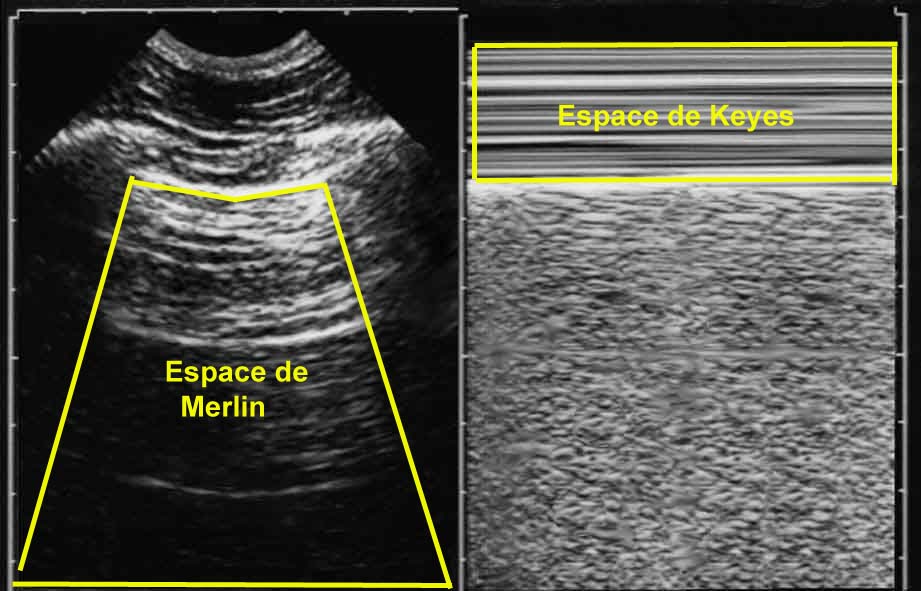
Our North-American colleague, Linda Keyes (Consulting Editor for Annals of Emergency Medicine & Associate Editor for Wilderness and Environmental Medicine) gave rise to the «Keyes space»: so was labelled this rectangular area, down limited by the pleural line, visible on a M-mode lung ultrasound image (1).
Christophe Perrin, Chair of the Pulmonoly department at Cannes hospital, will introduce the concept of CEURF at the Collège des Pneumologues des Hôpitaux Généraux.
(1) Merlin’s space (from Elisabeth Merlin, CEURFeur of Neo-Caledonia) was this space limited, on a real time image, between pleural line, shadow of ribs, and low limit of image (in Merlin’s space are displayed A and B-lines). Now, the concept of a Keyes space allows recognition, on a static view, of a dyspneic (BLUE-protocol) or not (PINK-protocol of ARDS) state of a patient during the examination. Dyspnea creates perturbances in the Keyes space, because of accessory muscles work. Following lung ultrasound rules, static images are even more informative.