INFORMATION on critical ultrasound
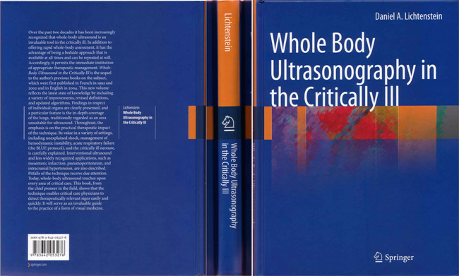
The textbook Whole Body Ultrasonography in the Critically Ill (2010) is just launched by Springer.
Since the first editions (1992, 2002 & 2005), this book received regular improvements. Critical ultrasound was
defined, in the 1992 Edition, as a circuit made from one ultrasound diagnosis and one immediate therapeutic
procedure, regarding a life-threatening situation. Lung ultrasound perfectly fulfills the definition of critical
ultrasound, also the main topic, since the BLUE-protocol, which combines lung and venous ultrasound for
immediate diagnosis of acute respiratory failure, is covered in 11 chapters. Pneumothorax, pleural effusion,
lung consolidation, phrenic assessment, and the relevance of the B-lines for diagnosing interstitial syndrome are
comprehensively detailed.
The BLUE-protocol is namely described in Chapter 20. The section on “Frequently Asked Questions about the
BLUE-protocol” will be of high interest for the practicionners.
Lung ultrasound characteristics in the neonate are dealt with in Chapter 21.
A simple approach to the heart is available in Chapter 22, barely unchanged since the 1992 Edition.
Chapter 23 includes the main novelty: the description of a new approach to acute circulatory failure. The
Limited Investigation (considering hemodynamic therapy) opens with a simple approach to the heart. It follows
with the FALLS-protocol, which is one application of lung ultrasound: Fluid Administration Limited by Lung
Sonography, i.e., mainly, the potential of detecting the ultrasound B-lines. The caval veins analysis then
follows, using an abdominal approach, but also a superficial approach for the superior caval vein.
Refinements are found in all chapters (the optic nerve and elevated intracranial pressure in Chapter 24, venous
access in Chapter 12, pneumoperitoneum in Chapter 5, mesenteric infarction in Chapter 6...). Chapter 13
introduces the BLUE-protocol, dealing with the peculiarities of a fully adapted technique for detecting deep
venous thrombosis in the critically ill.
Chapter 29 presents fast protocols for assessing the causes - and immediate therapies - of extreme circulatory
failure (the SESAME-protocol), assessing ARDS at the bedside, diagnosing pulmonary embolism in complex
ventilated patients with ARDS (the CLOT-protocol), finding the real cause of elevated temperature in
ventilated patients (the FEVER-protocol)....
In Chapter 30, notes about the material (in parallel with Chapter 2) are considered, explaining why, using the
approach described through the 330 pages, a simple material and a universal probe can be used without
compromise to the patient safety. Free talks are available regarding certain misconceptions in this recently
exploding field.
This textbook kept a minimal volume. Various settings are not specified (trauma, extreme environments,
spaceship, emergency room, involved disciplines such as pediatrics, cardiology, anaesthesiology, pulmonology,
thoracic surgery, radiology, family medicine...), since the applications, signs and material used are the same. A
single-author redaction was the key for an homogeneous amount of information.
Whole Body Ultrasonography in the Critically Ill, based on a 20-year experience in the field, opens the way of
a new discipline, for a simple approach to a genuine v
.
(Chapters highlighted in BLUE deal specifically with the BLUE-protocol)
Chapter 1 : Basic notions
Chapter 2: The ideal unit
Chapter 3: Specific notions to the critically ill in the ICU
Chapter 4: Basic anatomy
Chapter 5: Peritoneum
Chapter 6: GI tract
Chapter 7: Liver
Chapter 8: Gallbladder
Chapter 9: Urinary tract
Chapter 10: Miscellaneous abdominal organs
Chapter 11 : Aorta
Chapter 12: Central venous access
Chapter 13: Deep venous thrombosis adapted to the BLUE-protocol
Chapter 14: Lung, basic notions
Chapter 15: Pleural effusion
Chapter 16: Lung consolidation
Chapter 17: Interstitial syndrome
Chapter 18: Pneumothorax
Chapter 19: Lung ultrasound versus CT
Chapter 20: The BLUE-protocol
Chapter 21 : Lung ultrasound in the neonate
Chapter 22: Basic heart
Chapter 23: Limited Investigation considering hemodynamic therapy - the FALLS-protocol
Chapter 24: Head, optic nerve
Chapter 25: Soft tissues
Chapter 26: Interventional ultrasound
Chapter 27: Surgical ICU
Chapter 28: Outside the ICU
Chapter 29: Severe and frequent situations - algorithms
Chapter 30: Free considerations
Chapter 31: The training method of the CEURF
2010 Ed. 326 pages, 321 illustrations, 27 in color. 127 Euros. Hardcover. ISBN: 978-3-642-05327-6.
Order for reprints : Orders-HD-individuals@springer.com