Where to apply the probe: the BLUE-points
(standards exams points)
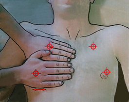
Two hands (of the size of that of the patient) are posed thus, starting from the clavicle. The medium of the higher hand determines the upper BLUE-point. Medium of the lower palm, the lower BLUE-point.
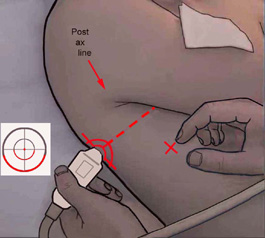
Leaving the lower BLUE-point transversely, a line crosses the posterior line axillaire. This crossing, or most subsequently possible, defines the PLAPS-point .
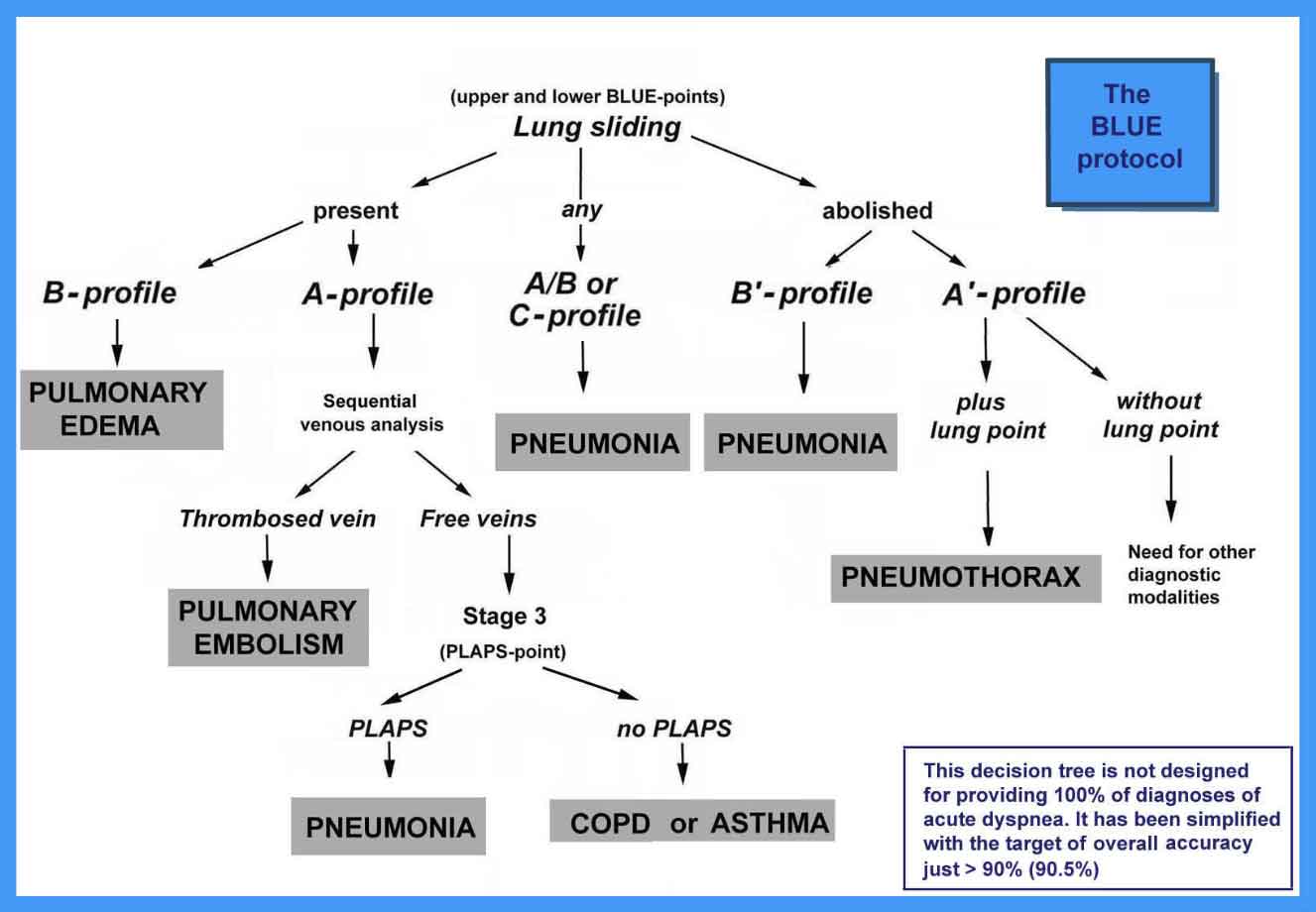
- A-profile (with DVT, usually pulmonary embolism)
- B-profile (usually hemodynamic pulmonary edema)
- B'-profile (usually pneumonia)
- A'-profile (usually pneumothorax)
- C-profile (usually pneumonia)
- A/B-profile (usually pneumonia)
- A-V-PLAPS profile (usually pneumonia)
- Nude profile (usually asthma and COPD)
- The original article: Relevance of lung ultrasound in the diagnosis of acute respiratory failure. The BLUE-protocol. Chest 2008;134:117-125
- The textbook: Whole Body Ultrasonography in the Critically Ill (Springer, 2010), page 194
- The textbook: Lung Ultrasound in the Critically III - the BLUE-protocol (Springer, 2016)